
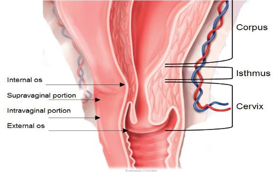
The name cervix derives from the Latin word “Cervic” meaning “neck”. It represents the lower portion of the uterus and communicates the uterine cavity with the vagina. It has a cylindrical shape with a length of about 3 cm and a diameter of about 2 cm. The uterine cervix has an opening to the vagina called the “external os” (EO). In the area of division between the cervix and uterine body lies a fibromuscular area called the “internal os” (IO). The area located between EO and IO is called the “endocervical canal”, which has a fusiform shape and an oval cross section, the endocervial canal has a diameter ranging between 3 and 10 millimiters.
The normal development of the uterus involves some changes happening over the mullerian ducts to form the cervix, uterus, fallopian tubes and upper vagina through the processes of differentiation, migration, fusión and canalization
The cervix has an intravaginal portion (portio vaginalis) and a supravaginal portion that is usually larger. Structurally, the cervix is composed of 85% extracellular matrix and 15% smooth muscle tissue. The extracellular matrix is made mainly of collagen, elastin and proteoglycans cells of smooth muscle and fibroblasts, epithelial cells and blood vessels.

Excelente mil gracias x tan enriquecedora información
LikeLike